 Image Database
NEURORADIOLOGY
BRAIN AND SKULL
Anatomy
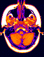
- Axial (SE T1W) MR images of brain
- Sagittal (SE T1W) MR images of brain
- Coronal (SE T1W) MR images of brain
- Whole Brain Atlas
(From the Department of Radiology, Brigham and Women's Hospital)
Pathology
- 7 hour Stroke: T2-weighted and Diffusion-weighted Imaging
(Courtesey of Dr. A. Gregory Sorensen and Dr. Bruce R. Rosen,
NMR Center, Massachusetts General Hospital, Boston, MA)
- 3 hour Stroke: T2-weighted and Diffusion-weighted Imaging
(Courtesey of Dr. Steven Warach, Beth Israel Hospital, Boston, MA)
- Craniopharyngioma (partly solid and partly cystic) - MRI
(download 40k JPEG image OR 134K GIF image)
- Bilateral cochlear otosclerosis (arrows: "double ring sign") - CT
- E-mail your answer for this case !! - MRI